Role: Design research, design of interactive learning games, 3D rendering and surface painting, graphic design, illustration, storyboarding, academic presentations
Project themes: 3D models, clinical imaging, pediatric critical care medicine
Timeline: January 2019 - January 2020
Partners: Dr. Charles Larson (pediatric intensivist), Cody Wesley, Patrick Von Hauff,
Thomas Jeffery
Thomas Jeffery
Photograph of Dr. Charles Larson sketching hypoplastic left heart syndrome by Cody Wesley
Pediatric Cardiac Intensive Care
Congenital heart defects (CHD) affect one percent of live births and are notoriously difficult to understand in their 3-dimensional (3D) form. Current educational practice for residents (junior doctors) leans heavily on memorization and diagrammatic representations of CHD. Patient education for visualizing what congenital heart defects look like relies on 2D illustrations, sourced from Google. There is a gap in the availability of anatomically accurate learning tools to understand CHD. This presented an opportunity to develop and document the design of a curriculum focusing on concrete forms of visualization.
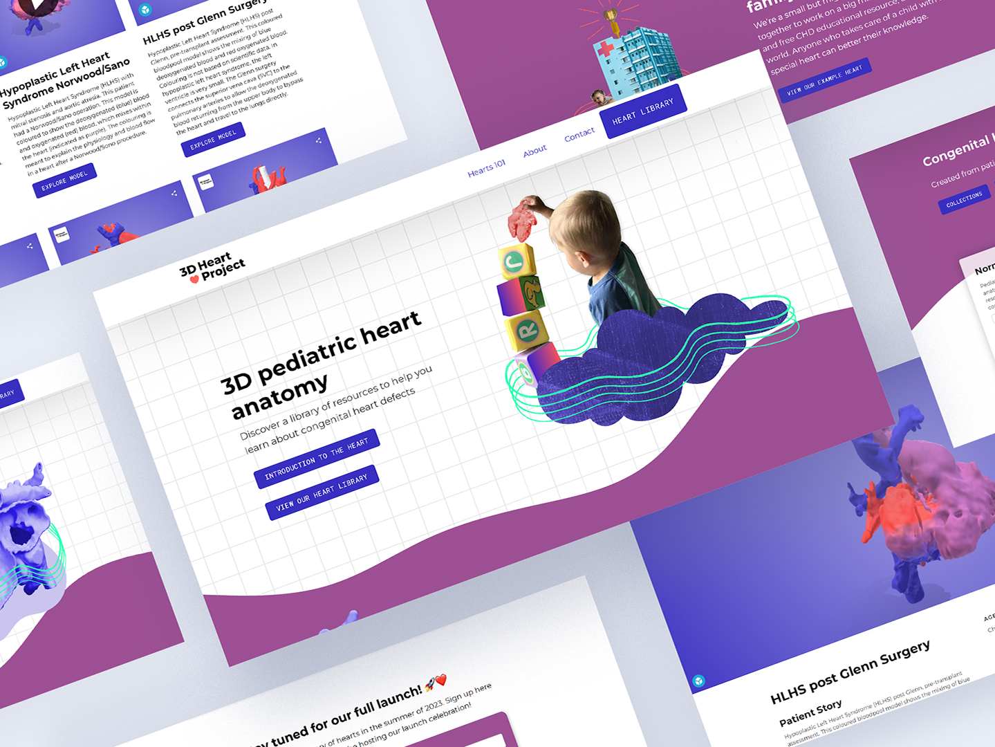
My role on this project was initially as a design practicum student, partnered with Cody Wesley, in the final year of my Bachelor of Design. The project has now evolved to become The 3D Heart Project, and my role continued into January 2020 as a Junior Designer at Academic Technologies.
Design Research
Resident doctors, who are doctors in training, are learners who are working on the hospital ward to gain experiential knowledge. They are active members of the care team, and experience many challenges on their way to specializing in a certain area of medicine.
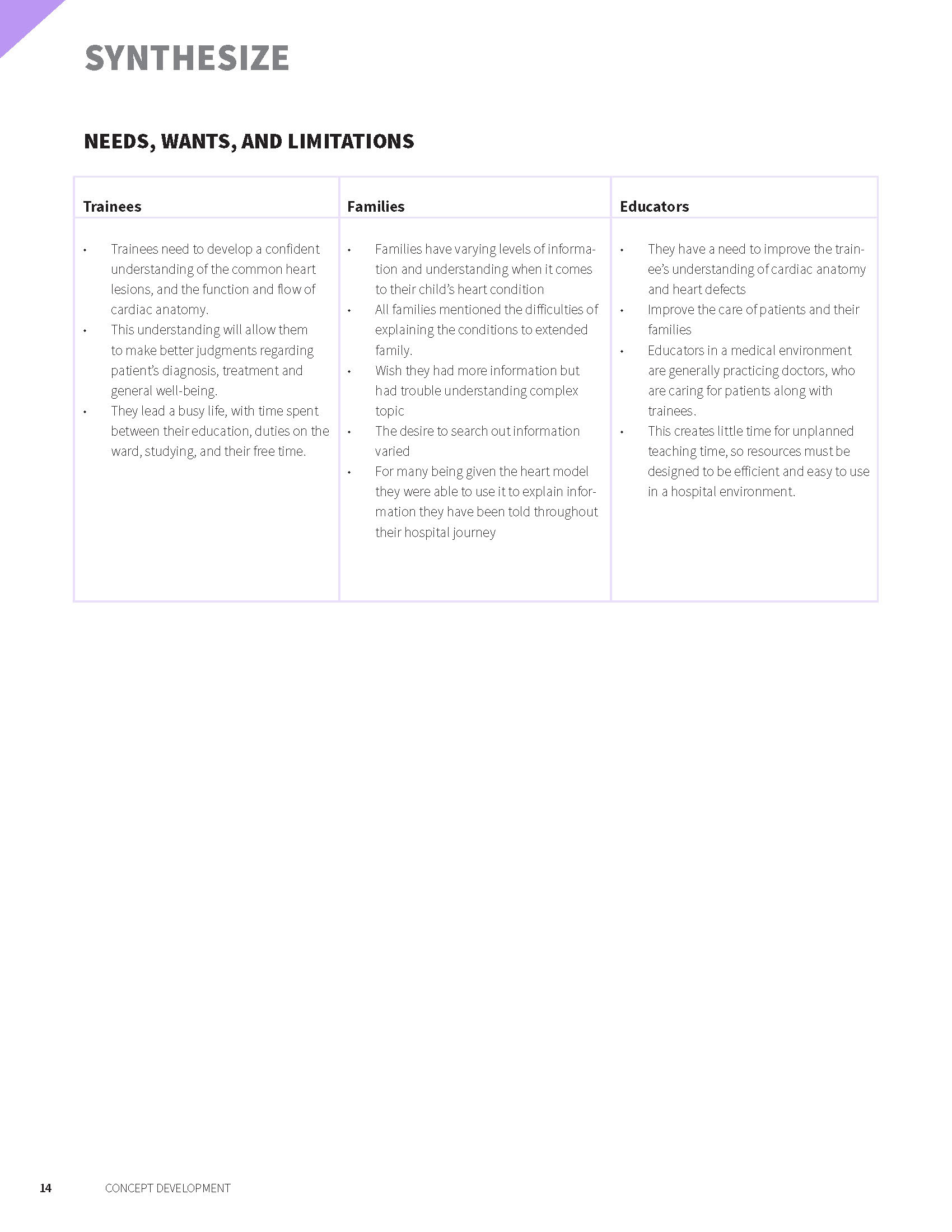
In our early design research, we needed to understand their lives, how they learned, their frustrations, and their desires for creating positive change in their education.
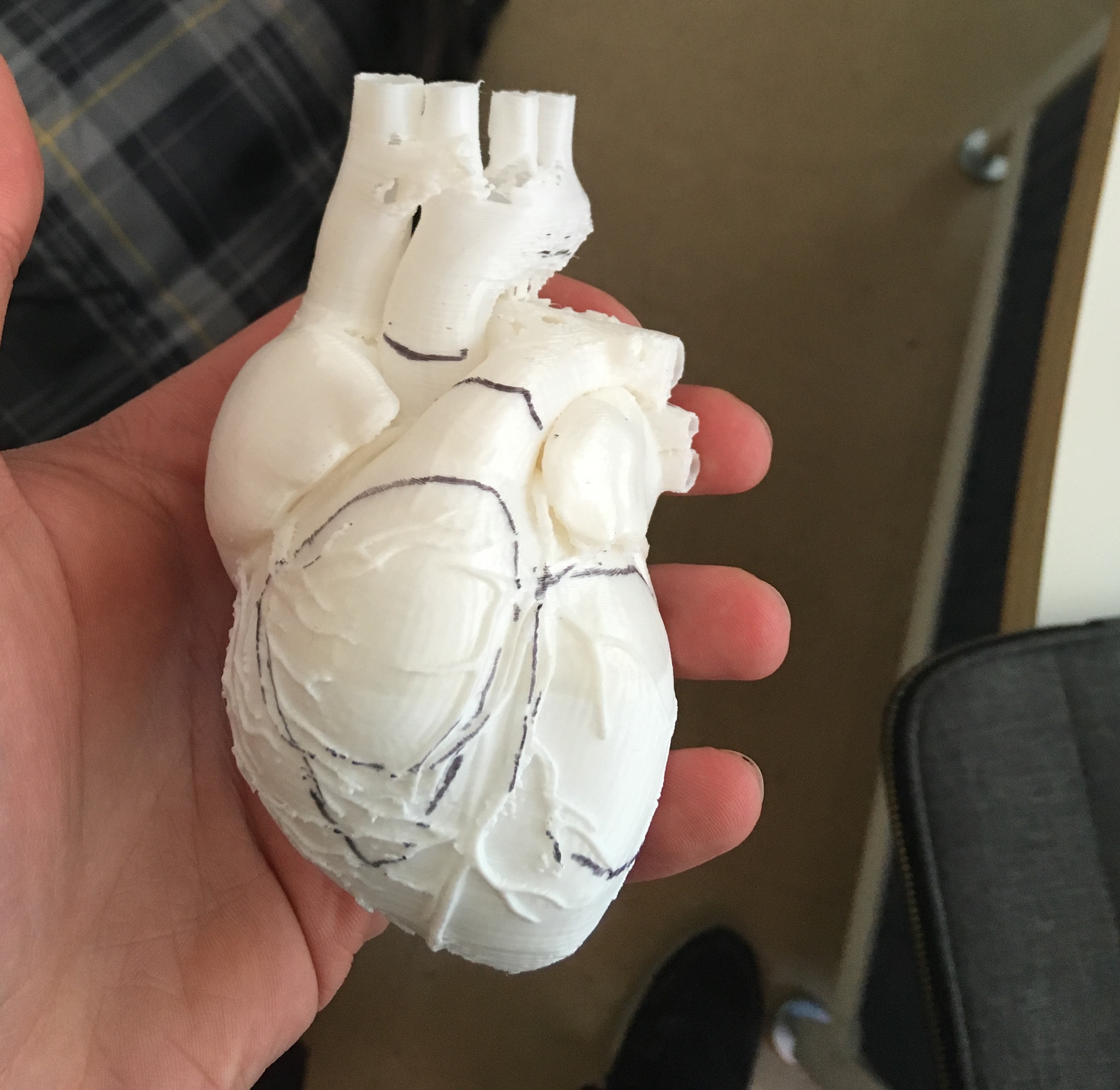
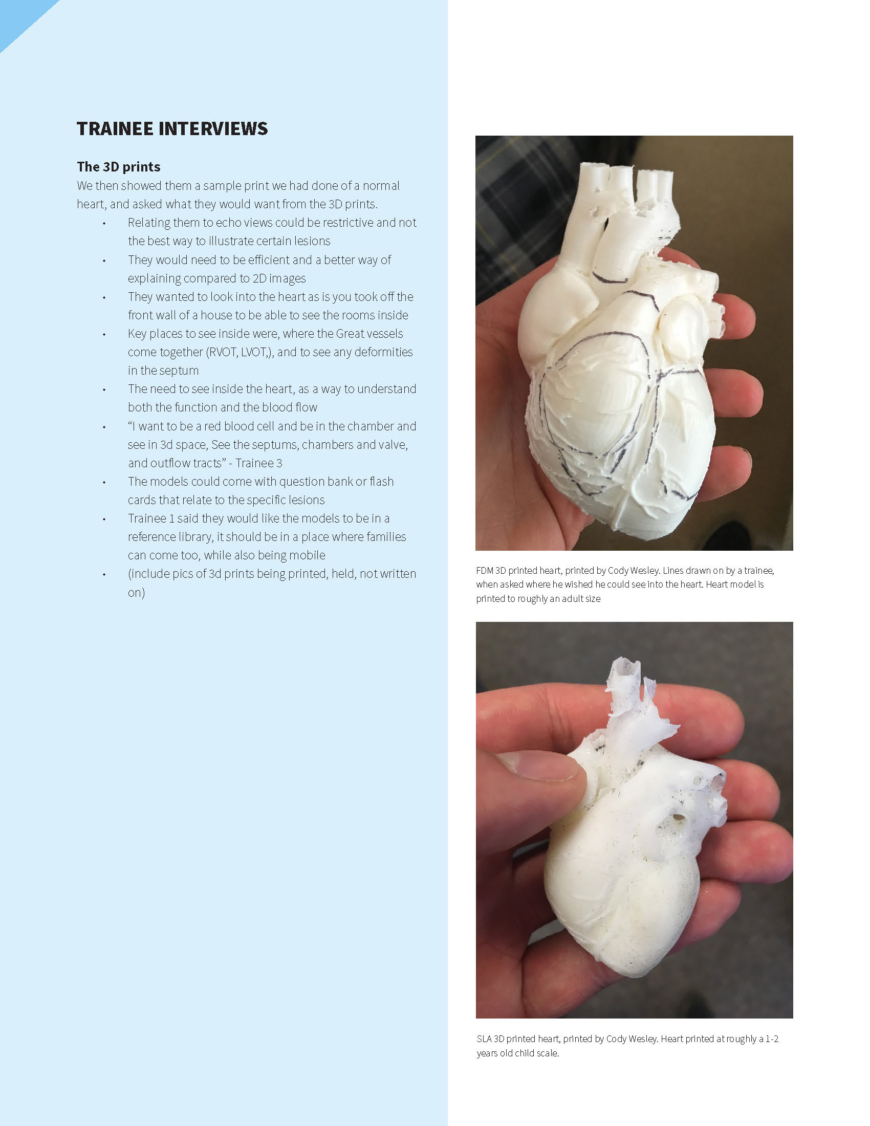
An early 3D print taken from a free 3D model online. We used this as an object to interview residents to ask where they wish they could see into the heart.
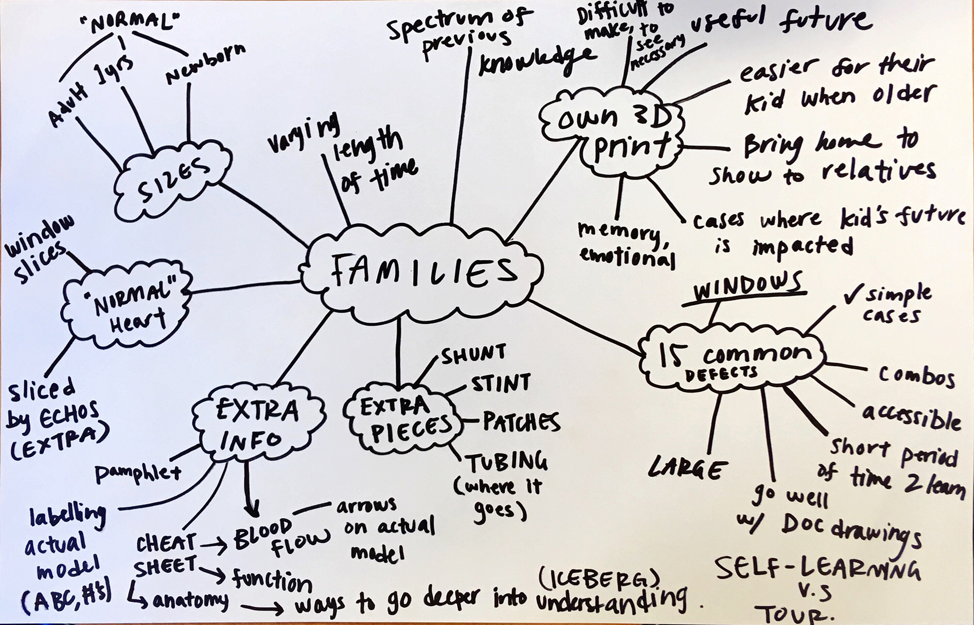
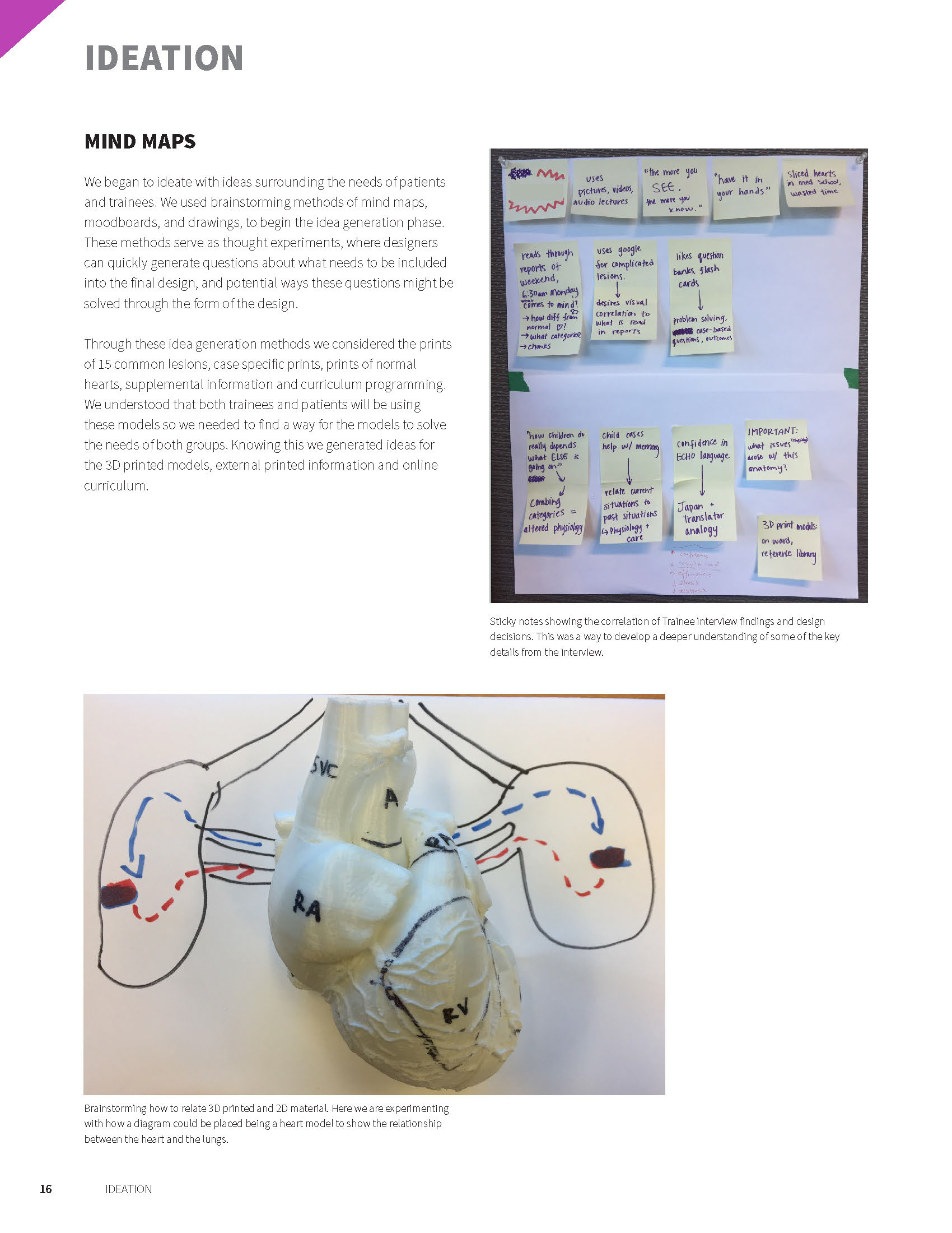
Mindmapping the needs, wants, and desires for 3D printed hearts for families.
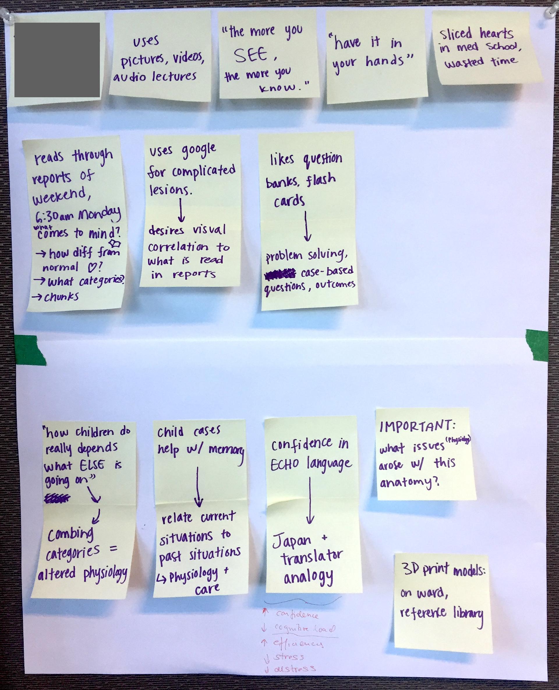
"Downloading our learnings" from an interview with a PCICU resident.
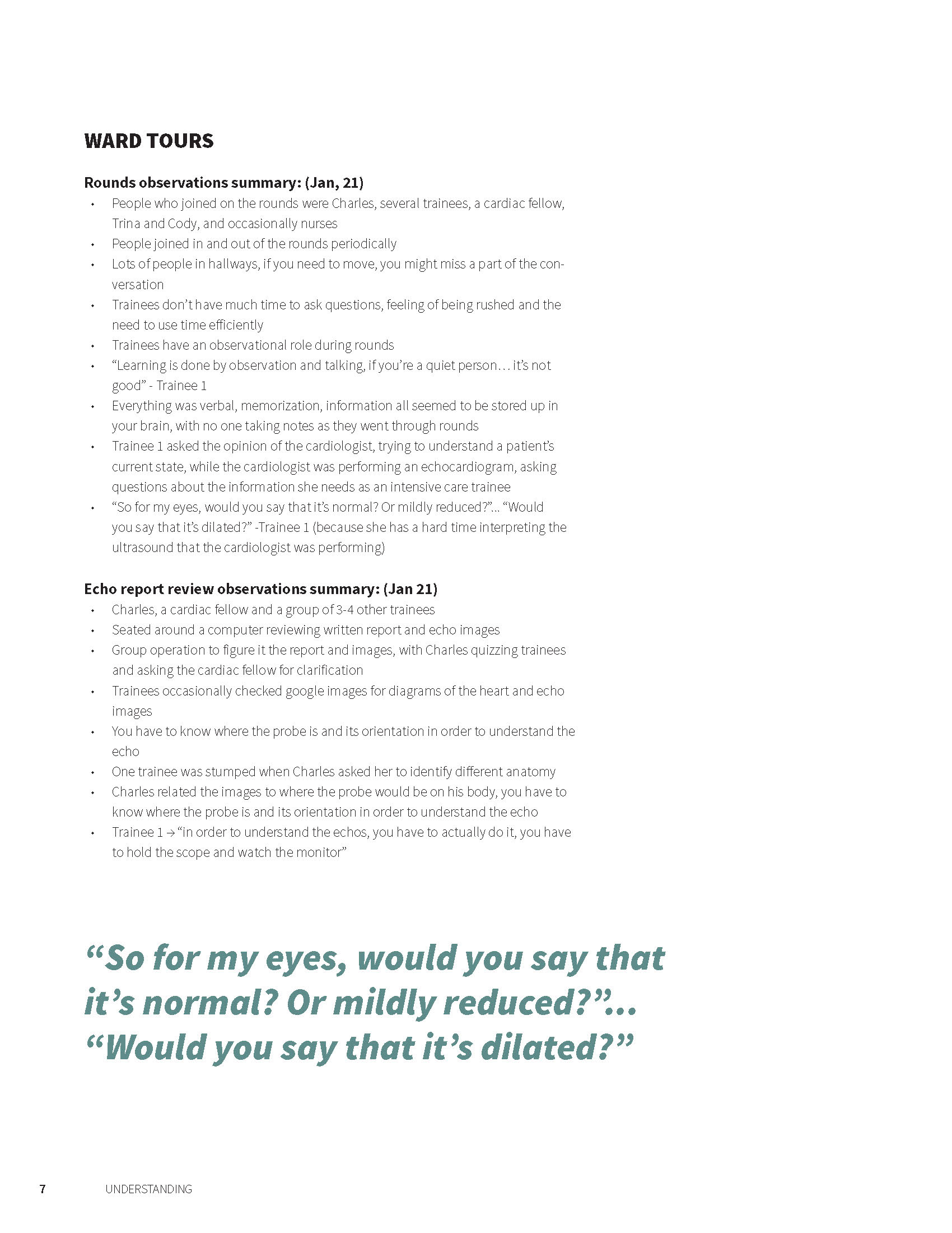
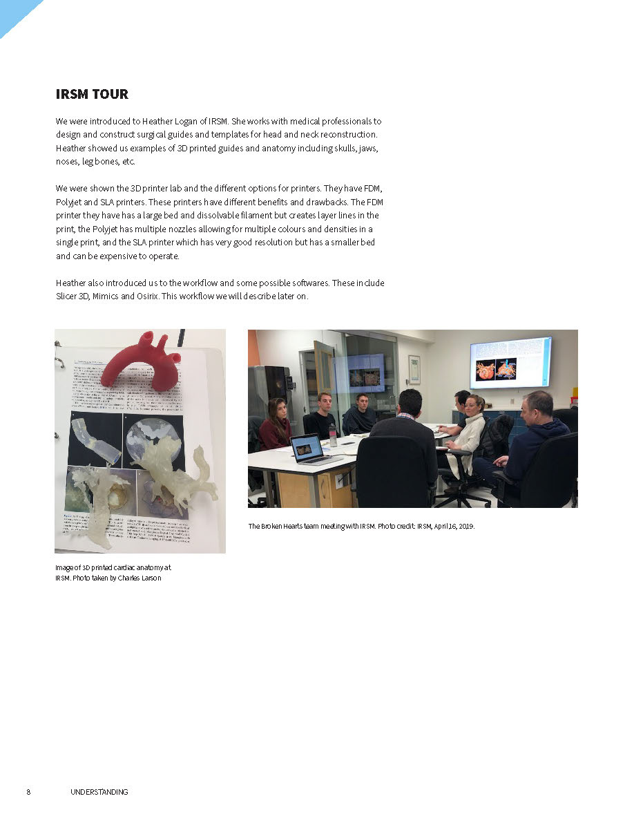
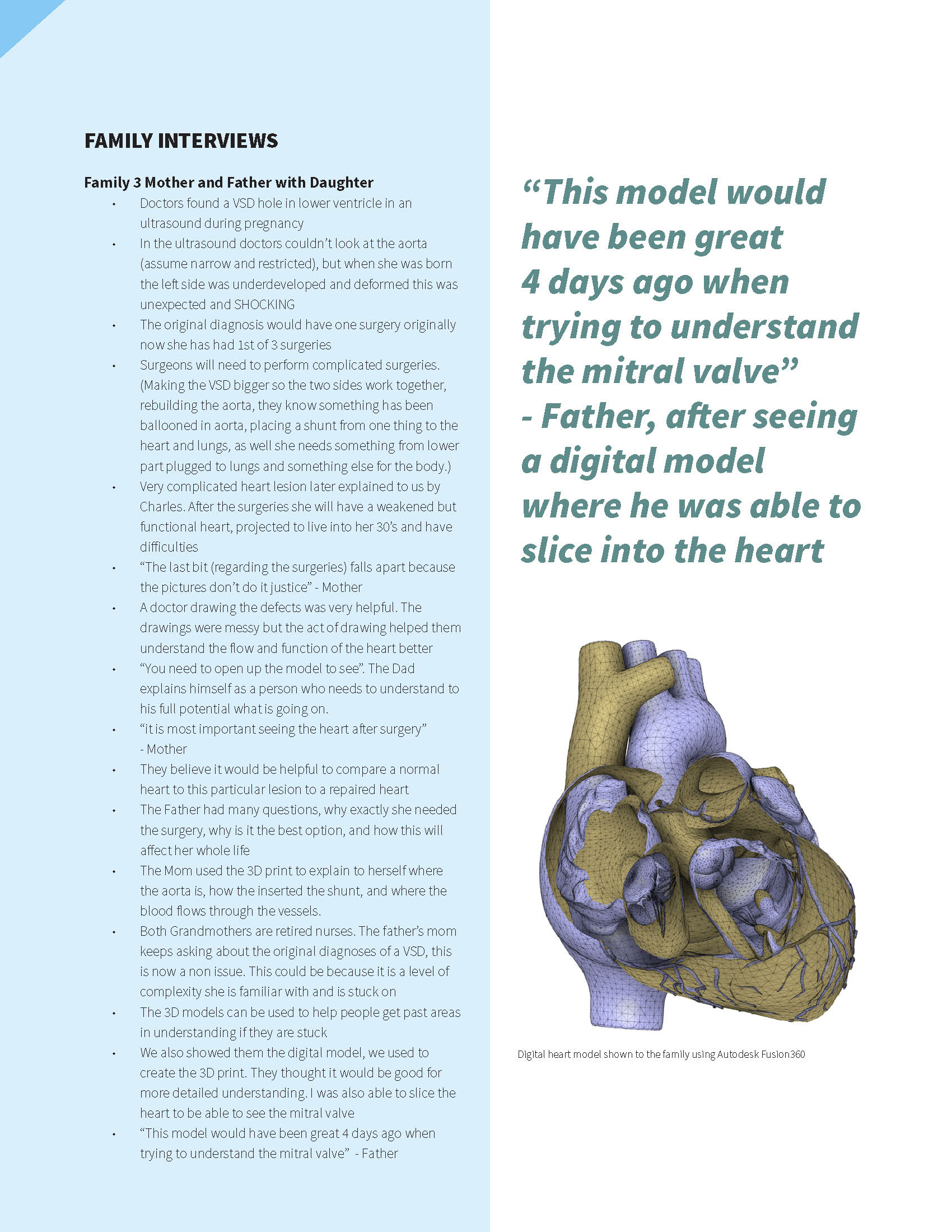
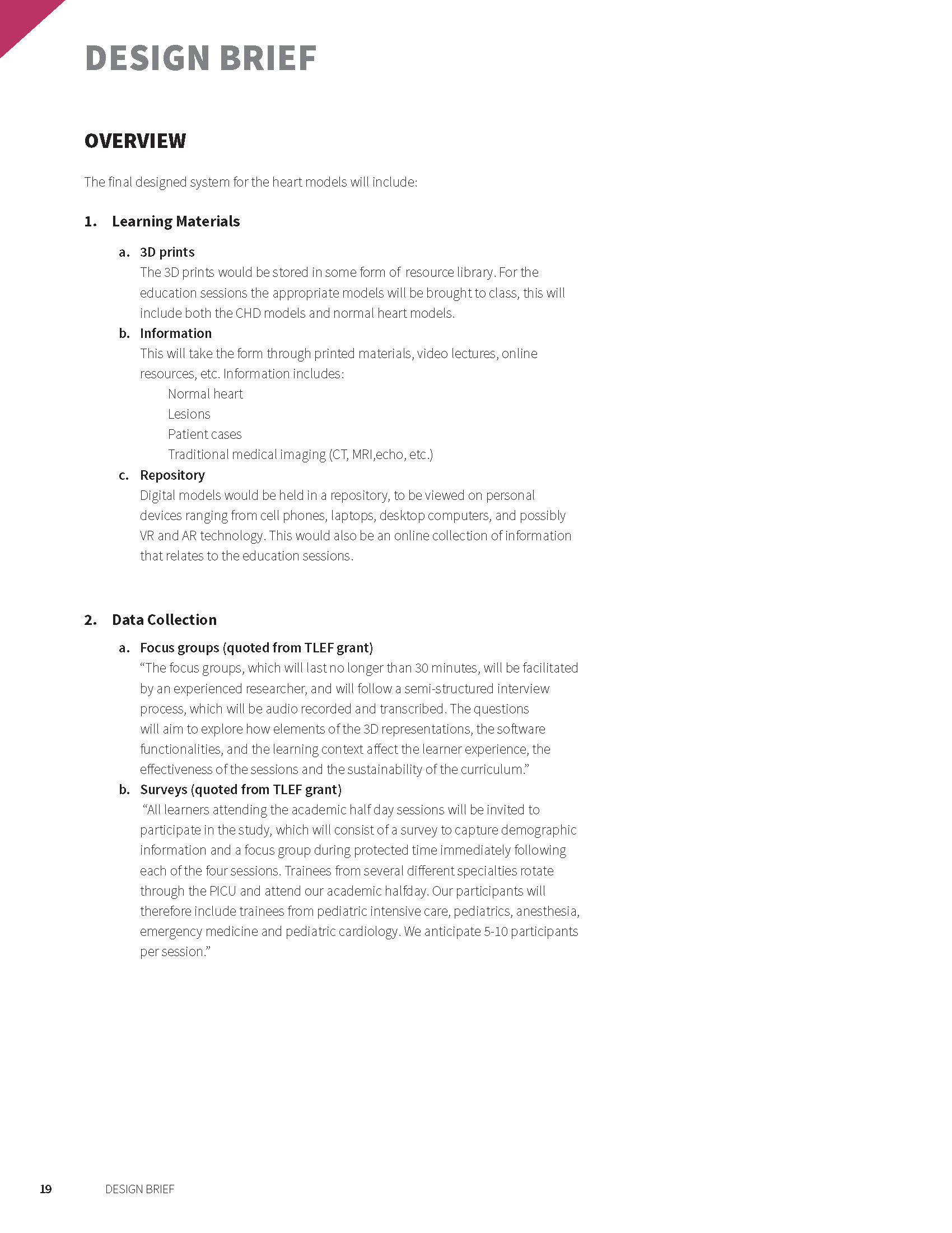
A booklet was created from our practicum to capture our findings from our own secondary research, tours, and interviews with trainees and families involved with CHD. We then synthesized these findings into a design brief, outlining how the 3D-printed heart models should be made and how the curriculum should be designed.
Ideation & Prototyping
We created ideas for how the models would be ideally created for 15 common congenital heart lesions. Each lesion would need specific care taken into the way it was represented in the 3D printed model, as the lesions manifest in varying anatomical structures within the heart.
A thought experiment investigating the question— how might we visualize an echocardiography scan as a 3D beam? A sketch model of a visual aid to communicate the sweeping motion of a 3D ultrasound scan.
An early prototype for a visual-spatial learning exercise for practicing spatial orientation, spatial visualization, and mental interpolation. This cube instructs resident learners to create a "cubed" model of a 3D-rendered congenital heart, and to diagnose the patient's defect. 3D heart model created by Cody Wesley.
Final Educational Materials
For the curriculum for resident learners, our final materials included 3D printed patient scans, "visuospatial ability cubes" (which are demonstrated in a video below), and an example of an information study sheet with 3D coloured renders. We tested these materials early on in August 2020 with resident learners, prior to using them in academic half day sessions later on in early 2020.
Dr. Charles Larson demonstrating the spatial ability cubes. A user receives a package of 6 images from different angles of a 3D heart, and must assign these images to the correct side of a cube. This exercise helps learners to identify and orient anatomical structures.
A flip-book sketch model of axial images of a CT scan of the heart, showcasing a congenital heart defect, to be used in conjunction with didactic learning (right object).
Research Poster
As a part of being a research assistant, I created a research poster for the Health Professions Education Student Research Presentation Day.
Telus World of Science, Dark Matters Night
Exhibition booth created in collaboration with Cody Wesley. January 23rd, 2020.
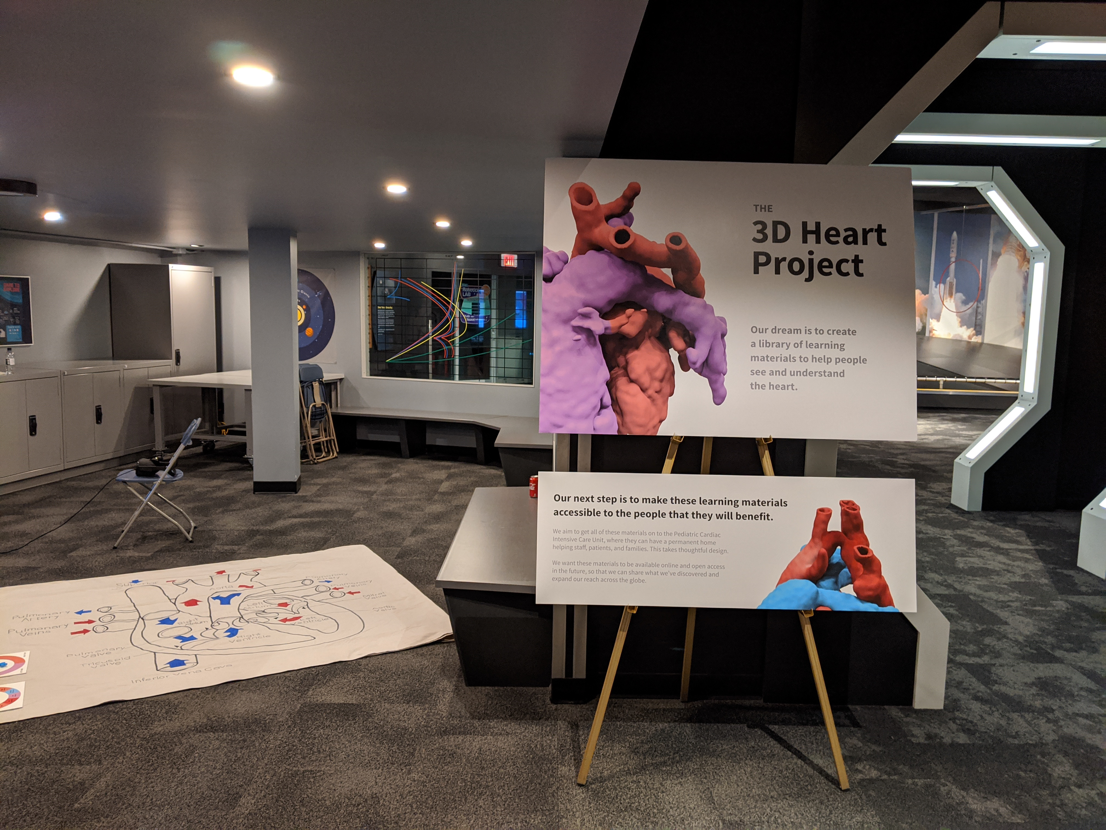
The 3D Heart Project at Telus World of Science event, Dark Matters.
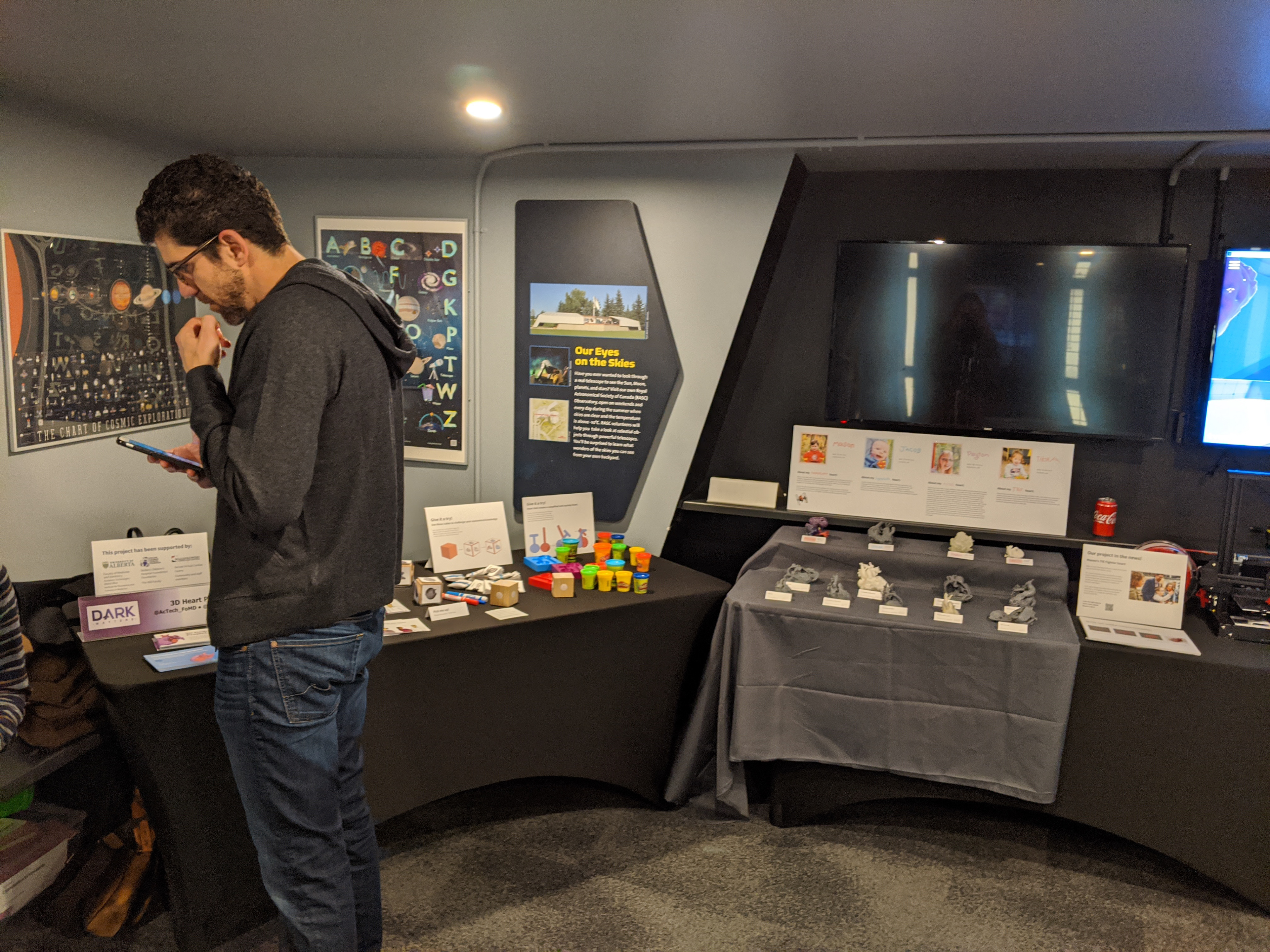
Dr Charles Larson with our displays featuring 3D printing, Heart-Doh, spatial cubes, and storybooks.
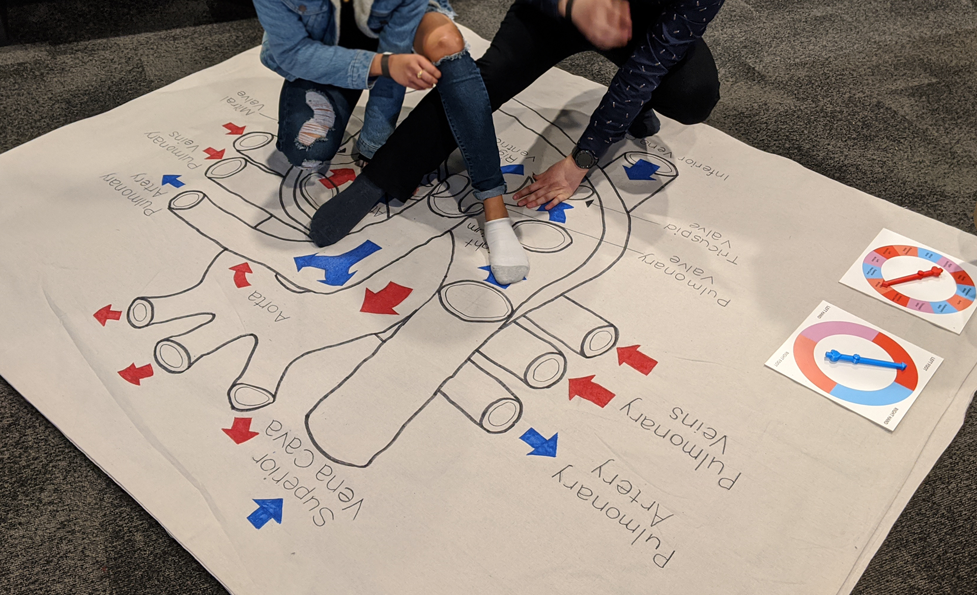
Heart Twister!!
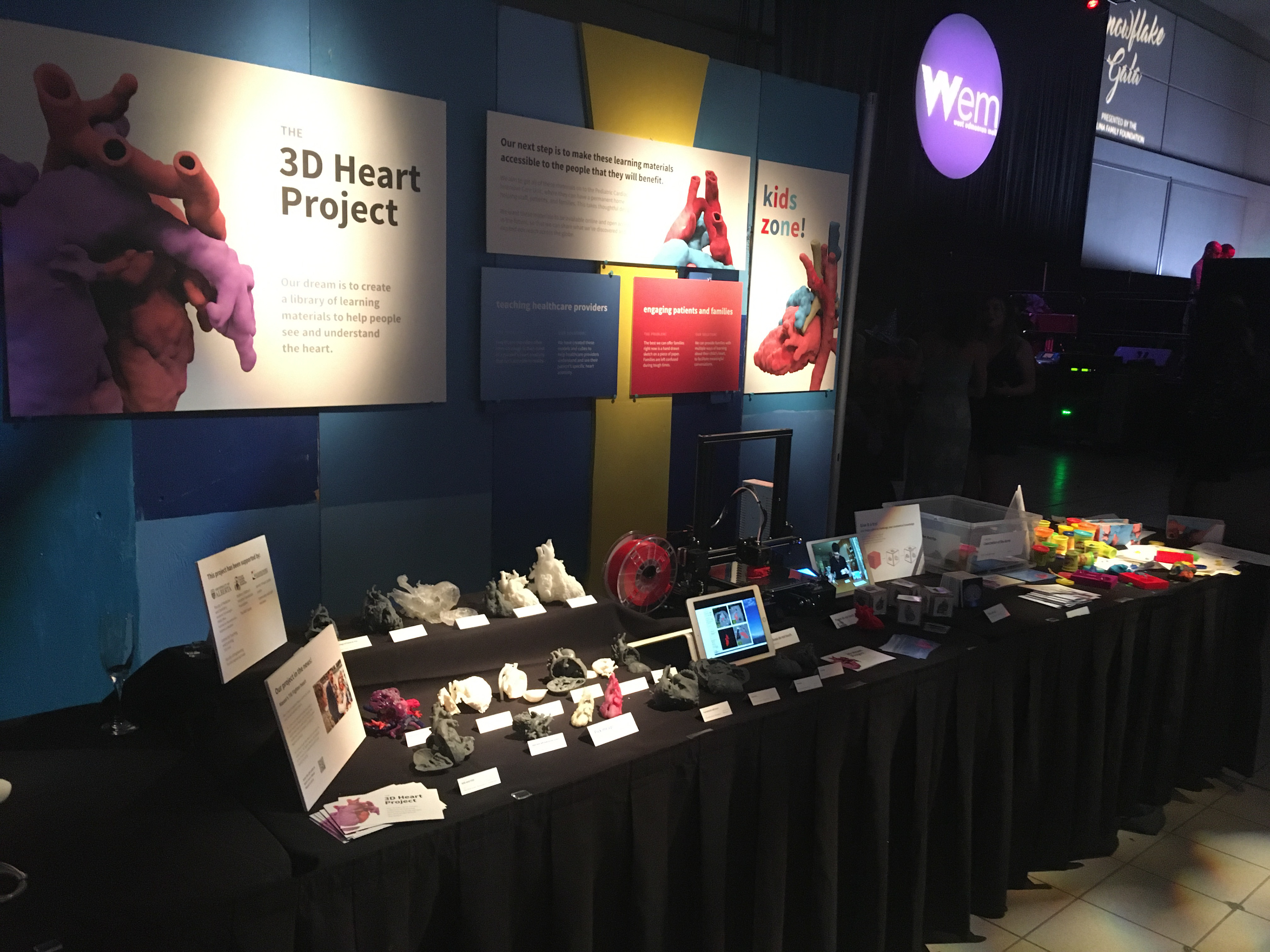
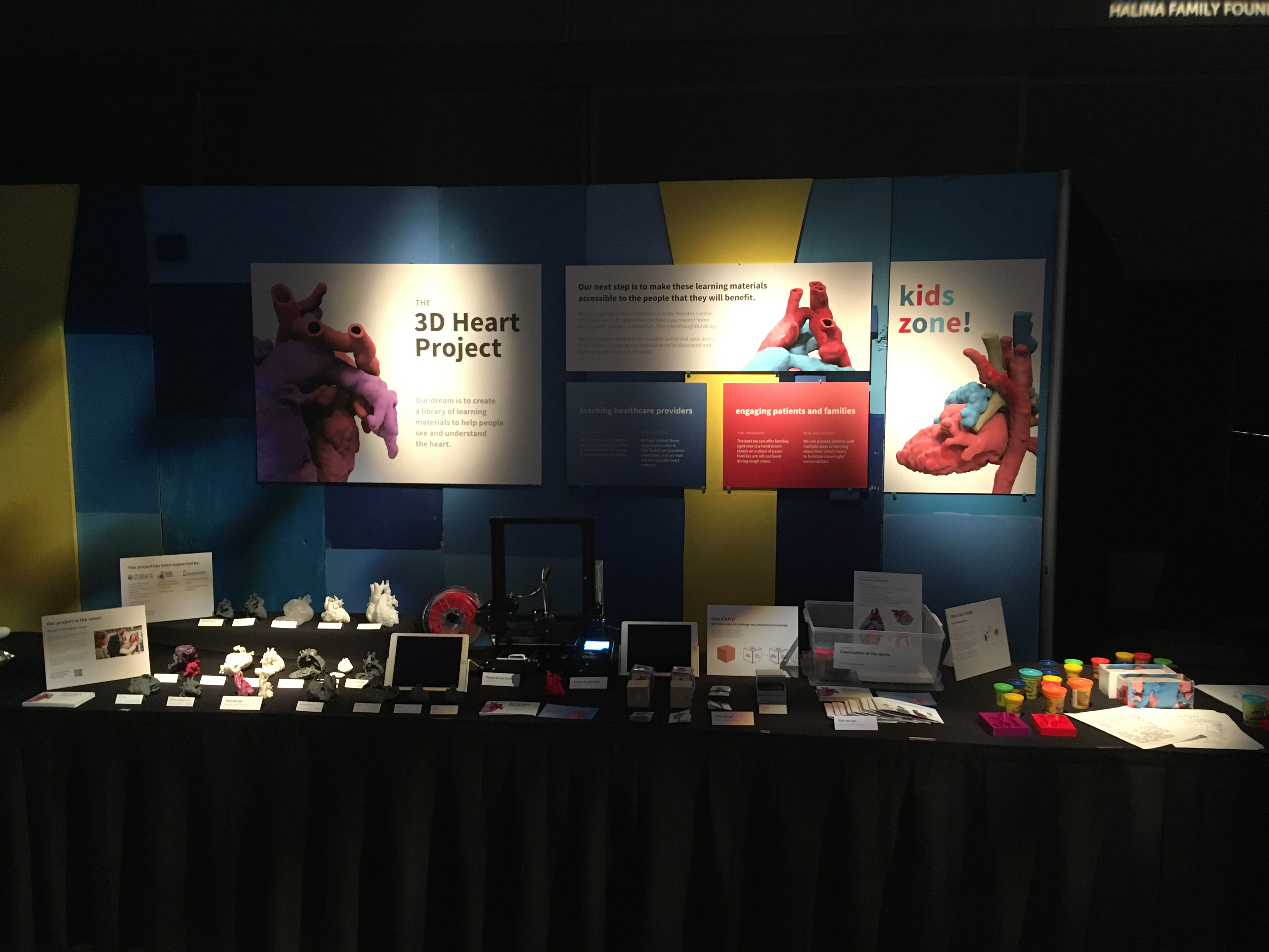
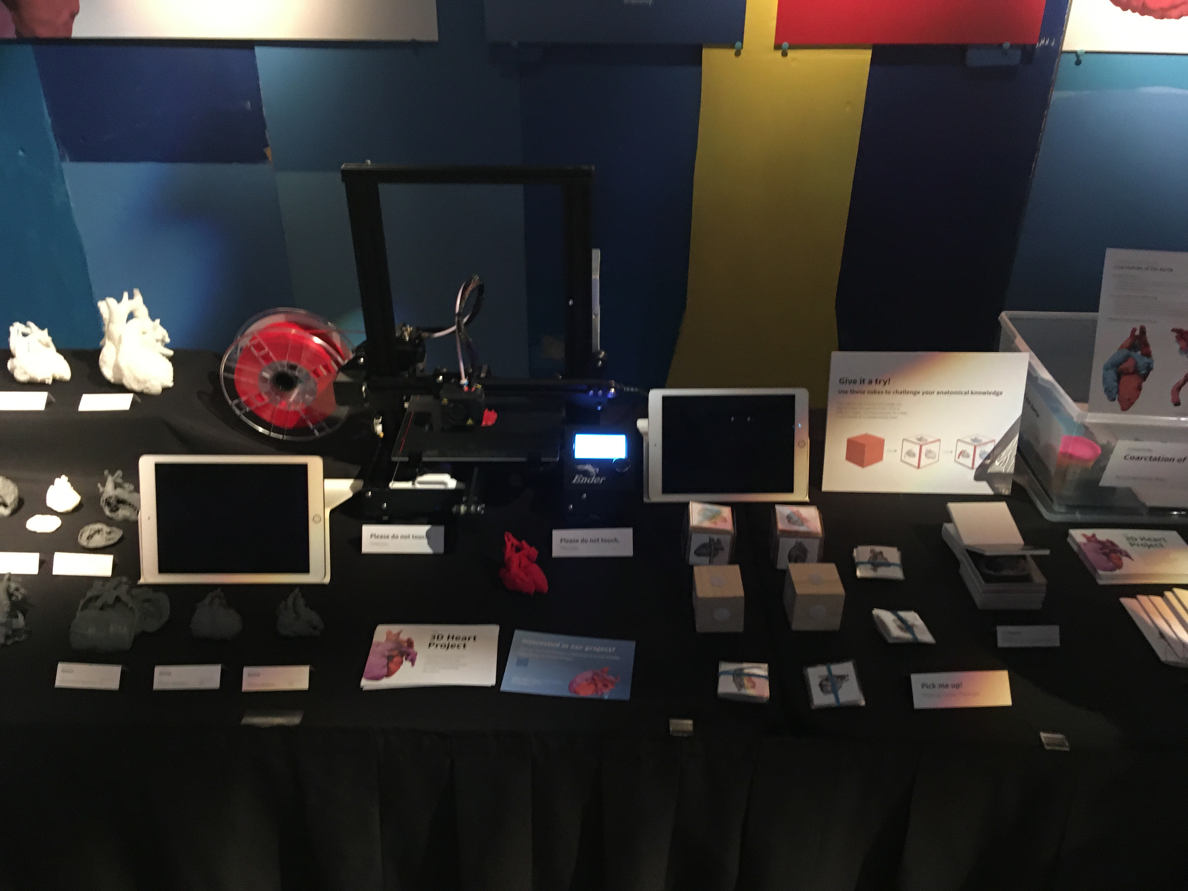
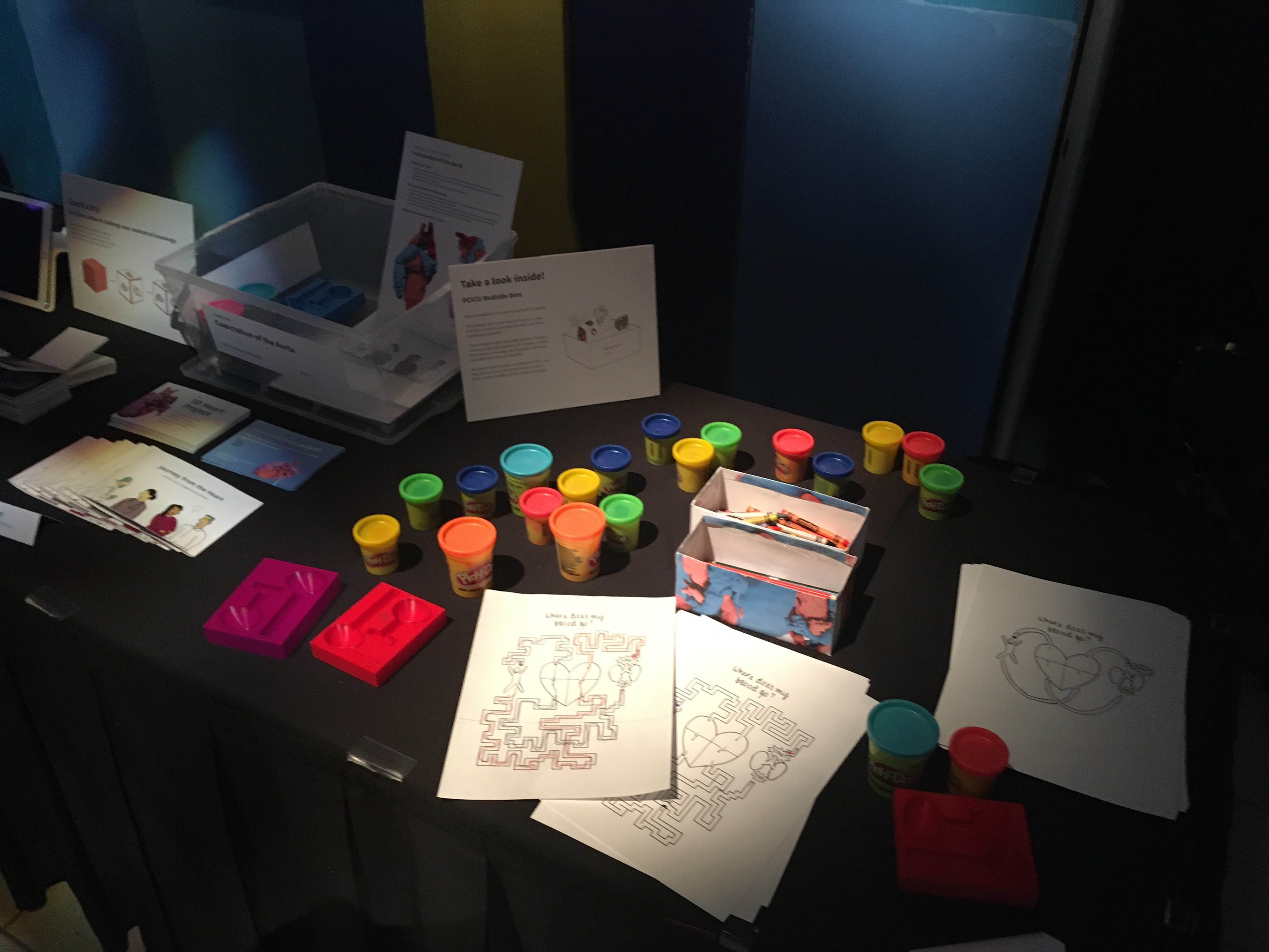
The Stollery Children's Hospital Snowflake Gala
Exhibition booth created in collaboration with Cody Wesley. December 2020.
